
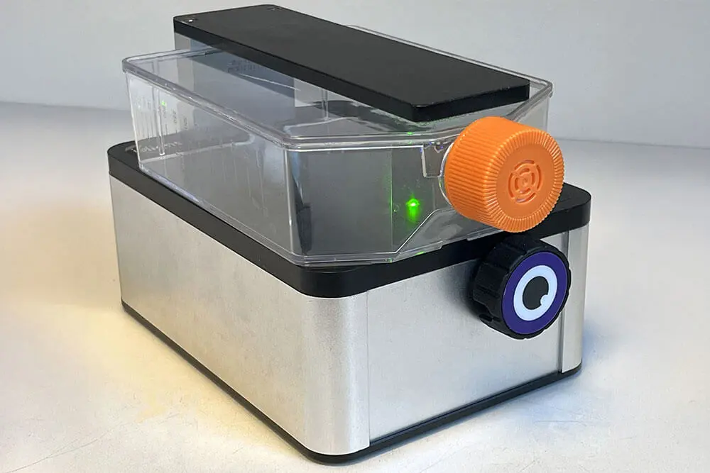
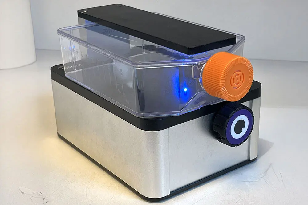

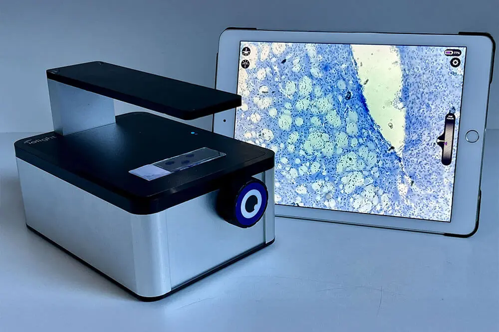
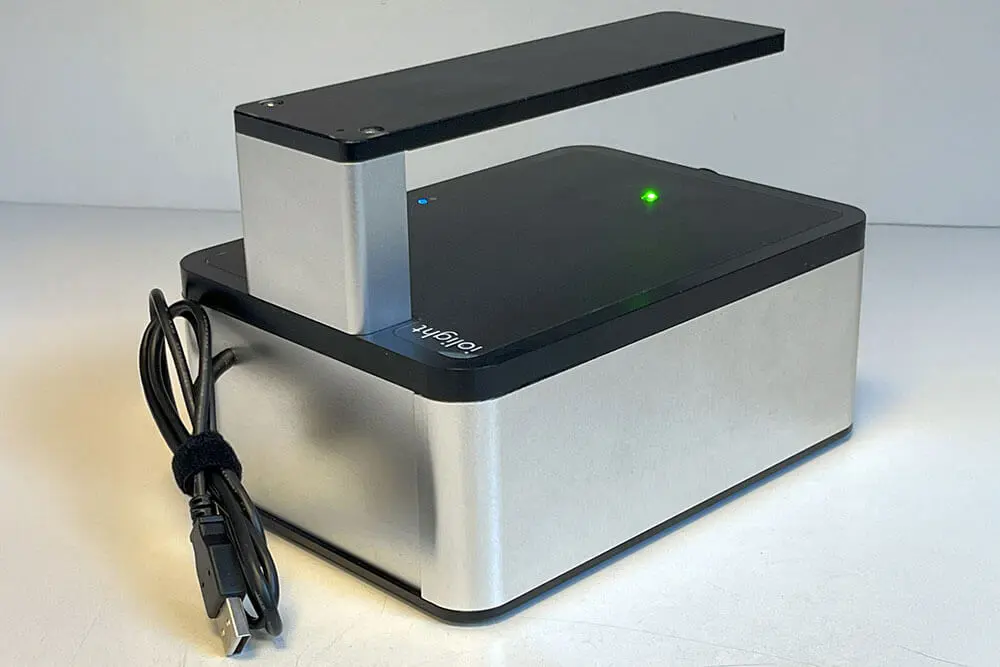
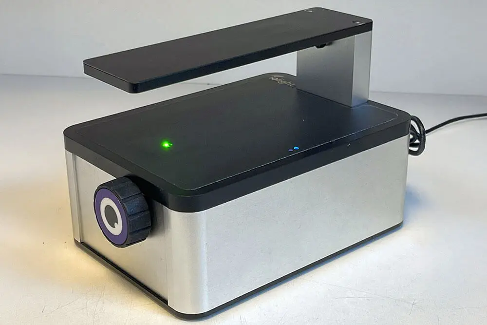



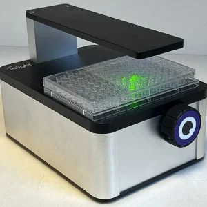
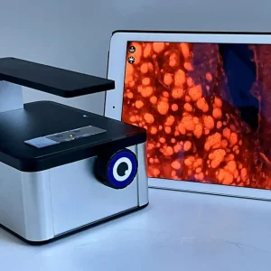
Compact Cell Imager with Dual Color Fluorescence
1mm field of view
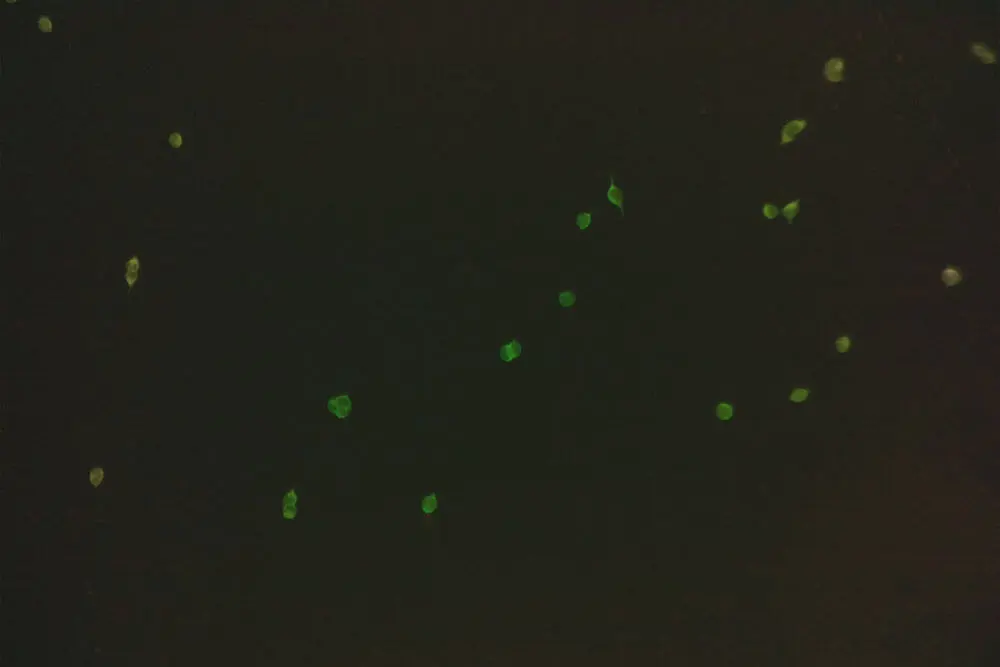
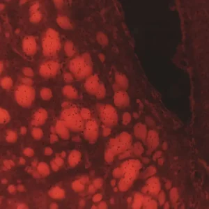
- Fluorescence: Blue and Green excitation
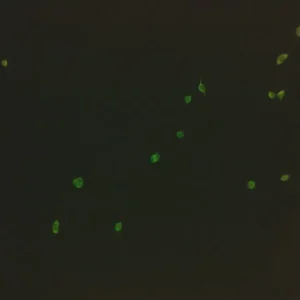
- Fluorophores GFP, YFP, PI, SYTO9, DS Red, tDTomato, Alexa Fluor, FITC, mCherry and more
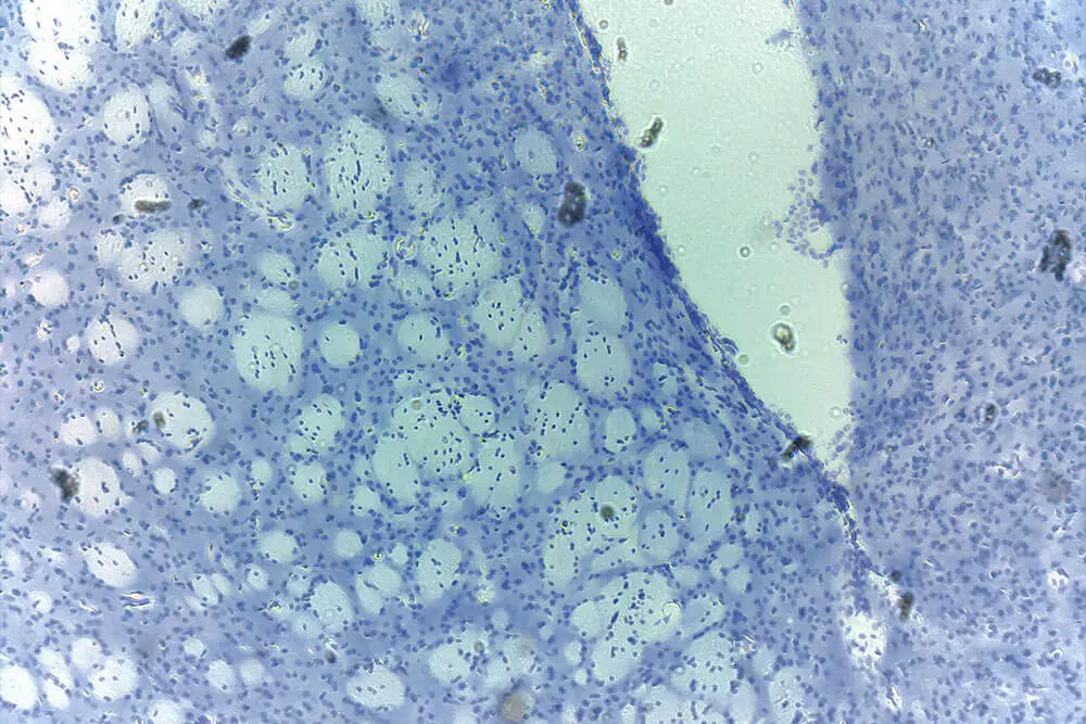
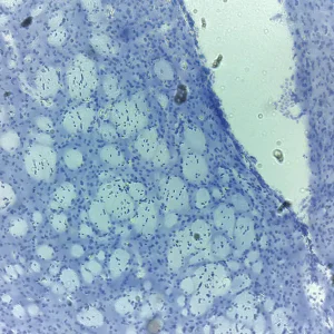
- Brightfield , darkfield and digital phase contrast imaging
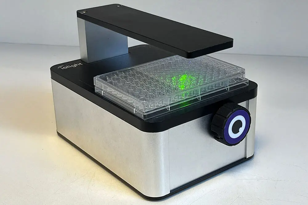
- Views samples from below in flasks, well plates and Petri dishes or on slides
- Can be left inside a CO2 incubator or safety cabinet and sterilised
- 1 micron resolution for viewing mammalian cells
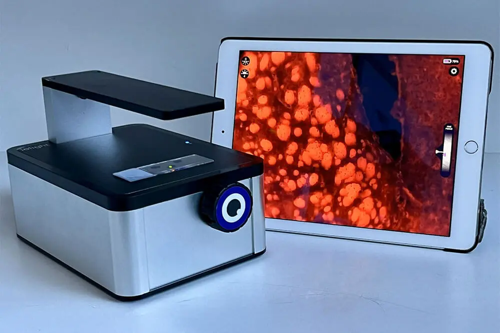
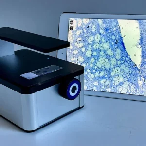
- Display images on a phone, tablet or computer without opening the door
- Instant sharing with colleagues anywhere
- Fast and simple to use
- Time-Lapse, Z-Stack, Z-stack combined with time-lapse and remote view
$14,795.00
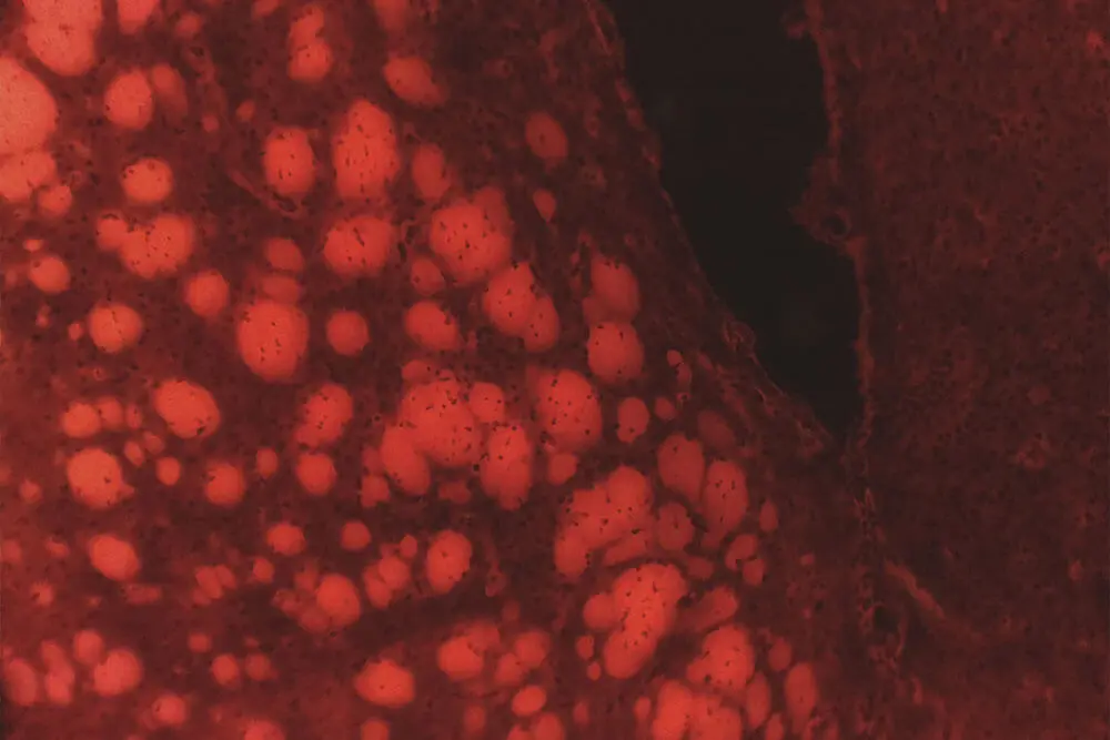
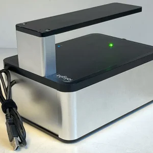
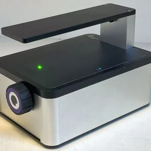
The ioLight compact inverted microscope is a sealed unit, whose small size allows for easy sterilisation and long term use in incubators or biosafety cabinets. The inverted microscope provides brightfield, darkfield and digital phase contrast imaging along with the capability to carry out timelapse or z-stack imaging. The ioLight compact cell imager with dual color fluorescence adds to this the capability of detecting common experimental fluorophores (GFP, YFP, PI Propidium Iodide, SYTO9, DS Red) tDTomato, Alexa Fluor, FITC, mCherry and more)
Incubator microscopes allow for continuous monitoring of cell cultures, such as HEK-293, CHO or HELA cells, and provide insights into cell numbers, cell morphology, cell health and identification of contaminants, increasing the number of successful cultures. This is key for small to medium scale production of bio-therapeutics, like stem cells for personalised medicine, with fluorescent markers potentially being used to follow stem cell differentiation or for quantification of transfection in the production of therapeutics. Furthermore the use of an incubator microscope allows for experiments such as wound healing assays or chemotaxis can also be carried out directly in the incubator, providing a stable and contaminant free environment for long running assays or delicate 3D cultures such as organoids.
Specifications
| Epifluorescence | Excitation switchable between 475nm or 560nm.Emission simultaneously detected at 515-535nm and 587-618nm by colour camera. Fixed lens and filter cubes so that the microscope is sealed for use inside incubators or safety cabinets. Easy sterilisation and cleaningEpifluorescence: Camera and Fluorescence illuminator are both below the sample |
|---|---|
| Field of View | 1.2 mm x 0.9 mm |
| Resolution | 1 micron |
| Maximum Magnification | Equivalent to x10 objective, 0.25 NA |
| Working Distance | 0 to 3mm stage to sample. |
| Maximum Flask Height | 48mm, taller flasks on request |
| Illumination | Trans illumination modes: Brightfield; Low NA brightfield; High contrast brightfield; Darkfield and Digital phase contrast. |
| Display (not included) | Smartphone, tablet, or computer with WiFi Direct connection, no internet connection or router is required |
| Image Sharing | Images and videos are stored on the display device for easy sharing. Images can be viewed remotely in Google Photos or any image sharing software. Time-Lapse on Android devices. |
| ioLight App | Free to download from the Apple® or Google Play® app store Embedded web server connects to Google Chrome and most other browsers on any WiFi computer Microscope makes a WiFi hotspot, no router or infrastructure necessary |
| Image Format | 5MP (2592 x 1944 pixels); colour JPG. BMP and PNG available on the web browser |
| Video Format | MPEG-4; colour; up to 1296 x 972; 10 fps Not available on web server |
| Z-Stacking | Web server only: 12 images at different focus points May be stacked using Photoshop, ImageJ or other image software |
| Weight | 1.9kg |
| Dimensions | 210 x 140 x 132mm, Taller flask models are >132mm high |
| Power | Integrated Li-ion rechargeable battery, USB charger supplied. Power lead is sealed to prevent ingress of the atmosphere Battery life: 5 hours with constant streaming. |
Requirements
- A tablet or phone running iOS 8.1 or higher or Android® 4.4 or higher, or a computer with WiFi and Google Chrome (or another web browser)
- We recommend newer, faster devices. Older low-performance devices may work, but this is not guaranteed.
What's In The Box
- Your ioLight Microscope
- USB charger, international model
- Users’ guide and certificate of conformity
Time-Lapse for Android
- The standard Android app allows Android Devices to record a sequence of Time-Lapse images from your ioLight microscope automatically. The app records images at regular intervals of a few minutes to days.
- Time-Lapse is great to use with the Inverted Microscope to monitor the growth of cells and cultures in incubators.
Specifications
| App | Requires Android device Now included in the standard Android App from the Google Play Store |
|---|---|
| Minimum Interval | 20 seconds |
| Number of Images | Limited by the storage available on the Android device. Each image requires <1Mb of storage space |
| Usage | For long runs, connect your Android device to power keep the app running in the foreground. |
| Remote Access | The ioLight Internet Module connects your ioLight microscope to the internet and your Android tablet at the same time. This means that Time Lapse images can be shared anywhere in real-time using Google photos or any other photo-sharing app. This allows researchers to view their experiments from home overnight or at the weekends. |
