
Real-world use: The University of Birmingham’s Center for Accelerated Drug Redeployment – March 2017
At the University of Birmingham—ranked among the top bioscience research institutions in the UK—Dr. Farhat Khanim and Professor Chris Bunce are leading groundbreaking cancer research through The Centre for Accelerated Drug Redeployment. Their work focuses on testing new therapeutic approaches to encourage cancer cells to return to normal behavior or die naturally—a vital step toward more effective cancer treatments.
 Overcoming Field Research Challenges
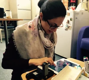
Much of Dr. Khanim’s work takes place in clinics located in remote areas, often without access to traditional laboratory facilities or trained lab staff. Conventional bench-top microscopes are too bulky, delicate, and complex for these environments.
The ioLight portable digital microscope provided an ideal solution—offering lab-quality imaging in a rugged, lightweight, and easy-to-use format.
With 1-micron resolution and built-in Wi-Fi connectivity, the ioLight enables researchers to capture and share high-resolution cell images instantly with colleagues back in Birmingham. This capability allows for real-time collaboration and faster decision-making—an essential advantage in research settings far from the lab.
Why Image Quality Matters
Understanding the life cycle of a cell is fundamental in cancer research. Healthy cells grow, perform specialized functions, and eventually die—a natural process called apoptosis. Cancer cells, however, fail to specialize and continue multiplying, leading to tumor growth.
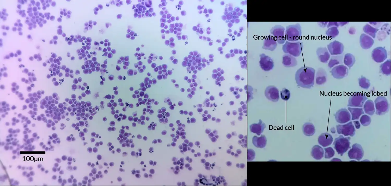
Dr. Khanim’s team studies how potential drug candidates might trigger cancer cells to “differentiate,” effectively resuming their normal life cycle and eventually dying off. This differentiation process can be identified visually under a microscope: as cells begin to change, their nuclei develop lobes and lose their smooth, circular shape.
Clear Results in the Field
Using the ioLight handheld digital field microscope, Dr. Khanim successfully captured high-quality images of leukemia cells, clearly showing nuclei beginning to develop lobes—an early indicator that the cells were responding to treatment and starting to differentiate.
These results demonstrated that ioLight’s portable microscope delivers the clarity and precision needed for advanced cancer research, even outside of traditional lab settings.
The ioLight advantage:
- 1 µm resolution for cellular-level imaging
- Portable and durable—ideal for field and clinical environments
- Wi-Fi connectivity for instant image sharing and remote collaboration
- Easy to operate, even by non-specialists